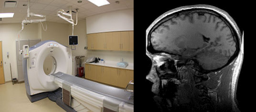
CT Scan/MRI Scan in India
What is CT scan ?
A CT scan makes use of computer-processed combinations of many X-ray images taken from different angles to produce cross-sectional (tomographic) images (virtual "slices") of specific areas of a scanned object, allowing the user to see inside the object without cutting. Other terms include computed axial tomography (CAT scan) and computer aided tomography.
Digital geometry processing is used to generate a three-dimensional image of the inside of the object from a large series of two-dimensional radiographic images taken around a single axis of rotation. Medical imaging is the most common application of X-ray CT. Its cross-sectional images are used for diagnostic and therapeutic purposes in various medical disciplines.The rest of this article discusses medical-imaging X-ray CT; industrial applications of X-ray CT are discussed at industrial computed tomography scanning.
Medical Use
Since its introduction in the 1970s, CT has become an important tool in medical imaging to supplement X-rays and medical ultrasonography. It has more recently been used for preventive medicine or screening for disease, for example CT colonography for people with a high risk of colon cancer, or full-motion heart scans for people with high risk of heart disease. A number of institutions offer full-body scans for the general population although this practice goes against the advice and official position of many professional organizations in the field.
Advantages
There are several advantages that CT has over traditional 2D medical radiography. First, CT completely eliminates the superimposition of images of structures outside the area of interest. Second, because of the inherent high-contrast resolution of CT, differences between tissues that differ in physical density by less than 1% can be distinguished. Finally, data from a single CT imaging procedure consisting of either multiple contiguous or one helical scan can be viewed as images in the axial, coronal, or sagittal planes, depending on the diagnostic task. This is referred to as multiplanar reformatted imaging.
What is MRI scan ?
Magnetic resonance imaging (MRI) is a medical imaging technique used in radiology to form pictures of the anatomy and the physiological processes of the body in both health and disease. MRI scanners use strong magnetic fields, radio waves, and field gradients to generate images of the organs in the body.
MRI does not involve x-rays, which distinguishes it from computed tomography (CT or CAT). While the hazards of x-rays are now well-controlled in most medical contexts, MRI can still be seen as superior to CT in this regard. MRI can often yield different diagnostic information compared with CT.
Medical uses
MRI has a wide range of applications in medical diagnosis and over 25,000 scanners are estimated to be in use worldwide.MRI affects diagnosis and treatment in many specialties although the effect on improved health outcomes is uncertain.Since MRI does not use any ionizing radiation, its use is generally favored in preference to CT when either modality could yield the same information.(In certain cases, MRI is not preferred as it can be more expensive, time-consuming, and claustrophobia-exacerbating).
CT/MRI
Procedure
o perform a study, the person is positioned within an MRI scanner that forms a strong magnetic field around the area to be imaged. In most medical applications, protons (hydrogen atoms) in tissues containing water molecules create a signal that is processed to form an image of the body. First, energy from an oscillating magnetic field is temporarily applied to the patient at the appropriate resonance frequency. The excited hydrogen atoms emit a radio frequency signal, which is measured by a receiving coil. The radio signal can be made to encode position information by varying the main magnetic field using gradient coils. As these coils are rapidly switched on and off they create the characteristic repetitive noise of an MRI scan. The contrast between different tissues is determined by the rate at which excited atoms return to the equilibrium state. Exogenous contrast agents may be given intravenously, orally or intra-articularly.
MRI requires a magnetic field that is both strong and uniform. The field strength of the magnet is measured in teslas – and while the majority of systems operate at 1.5 T, commercial systems are available between 0.2–7 T. Most clinical magnets are superconducting magnets, which require liquid helium. Lower field strengths can be achieved with permanent magnets, which are often used in "open" MRI scanners for claustrophobic patients
Diffusion MRI
Diffusion MRI measures the diffusion of water molecules in biological tissues.Clinically, diffusion MRI is useful for the diagnoses of conditions (e.g., stroke) or neurological disorders (e.g., multiple sclerosis), and helps better understand the connectivity of white matter axons in the central nervous system.