Retinal Detachment in India
About Retinal Detachment ?
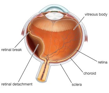
Retinal detachment is a disorder of the eye in which the retina peels away from its underlying layer of support tissue. Initial detachment may be localized or broad, but without rapid treatment the entire retina may detach, leading to vision loss and blindness. It is almost always classified as a medical emergency.
The retina is a thin layer of light sensitive tissue on the back wall of the eye. The optical system of the eye focuses light on the retina much like light is focused on the film or sensor in a camera. The retina translates that focused image into neural impulses and sends them to the brain via the optic nerve. Occasionally, posterior vitreous detachment, injury or trauma to the eye or head may cause a small tear in the retina. The tear allows vitreous fluid to seep through it under the retina, and peel it away like a bubble in wallpaper.
Signs and symptoms
fashes of light (photopsia) – very brief in the extreme peripheral (outside of center) part of vision.
a sudden dramatic increase in the number of floaters.
a ring of floaters or hairs just to the temporal (skull) side of the central vision.
Risk factors
Risk factors for retinal detachment include severe myopia, retinal tears, trauma, family history, as well as complications from cataract surgery.
Epidemiology
Retinal detachment is more common in people with severe myopia (above 5–6 diopters), in whom the retina is more thinly stretched. In such patients, lifetime risk rises to 1 in 20.About two-thirds of cases of retinal detachment occur in myopics.Myopic retinal detachment patients tend to be younger than non-myopic ones.
Retinal Detachment

Diagnosis
Slit Lamp Biomicroscopy Retinal Screening Programs: Systematic programs for the early detection of diabetic retinopathy using slit-lamp biomicroscopy. These exist either as a standalone scheme or as part of the Digital program (above) where the digital photograph was considered to lack enough clarity for detection and/or diagnosis of any retinal abnormality.
A scleral lens is a large contact lens that rests on the sclera and creates a liquid-filled vault over the cornea. In dry eye sufferers this lens bathes the cornea, reducing blurred vision caused by dry eye and providing relief from the dry eye pain caused by the sensitive nerves in that area.
Treatment
Find all retinal breaks
Seal all retinal breaks
Relieve present (and future) vitreoretinal traction